Fluorescent Labeled Oligonucleotides

Custom DNA and RNA dye labeling with direct synthesis or post-synthetic conjugation.

Custom fluorescent-labeled oligonucleotides for qPCR probes, FISH imaging, molecular diagnostics, hybridization, and multiplex fluorescence assays.

5′ / 3′ / Internal labeling Direct amidite or post-synthetic HPLC purified DNA & RNA compatible 200+ dye options

Overview

Bio-Synthesis provides custom fluorescent labeling of oligonucleotides for molecular diagnostics, quantitative PCR, fluorescence in situ hybridization (FISH), imaging, and multiplex detection assays. A broad range of fluorophores is available, including standard qPCR reporters, high-brightness imaging dyes, far-red and near-infrared labels for reduced autofluorescence, and dark quenchers used in dual-labeled probes, molecular beacons, and real-time detection systems.

Fluorescent labeled oligonucleotides are custom DNA or RNA constructs modified with reporter dyes for nucleic acid detection, molecular diagnostics, imaging, hybridization, and quantitative PCR workflows. Fluorophores can be introduced during solid-phase oligonucleotide synthesis using dye phosphoramidites or installed after synthesis through reactive handles such as amino, thiol, azide, alkyne, or DBCO groups, enabling flexible probe design for qPCR, FISH, molecular beacons, microarrays, sequencing, and advanced fluorescence-based assays [1–3].

Compared with unlabeled oligos, fluorescently modified oligonucleotides provide direct optical readout, multiplexing potential, and compatibility with quencher-based signal control. Careful selection of dye class, excitation/emission spacing, labeling position, spacer chemistry, and purification strategy is important for achieving strong signal-to-noise performance and reproducible assay behavior, especially in multiplex probe systems and imaging applications [4–6].

architecture-of-fluorescent-labeled-oligonucleotides overview schematic
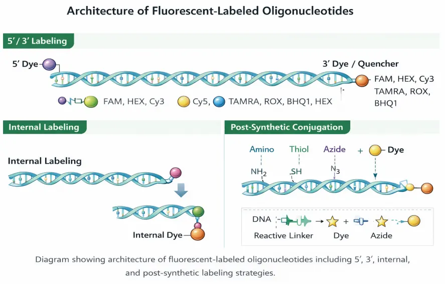
Architecture of Fluorescent-Labeled Oligonucleotides. Representative 5′ labeling, 3′ dye or quencher labeling, internal labeling, and post-synthetic conjugation strategies used for fluorescent probe and assay design.

Design insight: the best fluorescent labeling strategy depends on the assay format, optical channel plan, dye–quencher pairing, labeling position, and whether the fluorophore is better introduced during synthesis or through post-synthetic conjugation.

Fluorescent Labeling Positions

Position Description Common Uses
5′ End Dye attached at the 5′ terminus of the oligonucleotide. qPCR probes, sequencing, hybridization assays
3′ End Dye attached at the 3′ terminus. Reporter/quencher systems, FRET probes
Internal Dye incorporated at a defined internal nucleotide position. Molecular beacons, structure-sensitive assays
Dual Label Reporter + quencher or two-fluorophore design. TaqMan probes, FRET, multiplex probe systems

Labeling Chemistry

Direct amidite incorporation

Established fluorophores can be introduced during solid-phase oligo synthesis using dye phosphoramidites or dye supports. This is commonly preferred for standard reporter dyes and routine labeled probe formats.

Post-synthetic conjugation

Sensitive or custom dyes can be added after synthesis using reactive handles such as Amino C6, Amino TEG, thiol, azide, alkyne, or DBCO. This gives broader flexibility for specialty fluorophores and orthogonal conjugation strategies.

Spectral Dye Groups for Easier Channel Planning and Assay Design

This section keeps the cleaner service-page design of your second file while expanding the dye content using the richer by-color list from the first file. Each color family is now collapsible, and the section header keeps the circular color-dot styling for faster visual scanning.

Fluorescence Spectrum Guide

Grouped by color family to simplify multiplex planning and instrument-channel selection.

350 nm
400
450
500
550
600
650
700+
UV / Violet
Blue
Cyan
Green
Yellow
Orange
Red
Far-Red / NIR

Includes UV / violet labels plus the lighter weak-UV group from your source list.

Dye Excitation (nm) Emission (nm) Highlights
Pyrene 340 376 Excimer-forming dye with long fluorescence lifetime for hybridization studies
Alexa Fluor 350 345 444 Bright, photostable violet dye for multicolor applications
AMCA-X SE 350 450 Amine-reactive coumarin dye for protein and oligo labeling
DyLight 350 353 432 UV-excitable dye used in DNA labeling and microscopy
DyLight 405 400 421 Bright violet-blue dye used for multiplex imaging
Pacific Blue 410 455 Bright blue-violet dye used in flow cytometry and immunofluorescence
Pyridine Weak UV group component used as part of larger fluorescent scaffolds

Merged from Blue, Blue (Oxidized), Blue (Weak), Blue-Green, and Pale Blue groups in your first file to keep the second file cleaner while preserving the richer dye coverage.

Dye Excitation (nm) Emission (nm) Highlights
Marina Blue 365 460 Highly photostable dye for confocal and multi-color assays
Cascade Blue 377 423 UV-excitable dye with narrow emission
Alexa Fluor 405 401 421 Bright violet-blue dye with minimal overlap with green fluorophores
CF405S 404 431 Brighter than Alexa 405; good for SIM and 405 nm laser
ATTO 390 390 479 UV-excitable blue dye with low autofluorescence background
ATTO 425 425 484 Blue-violet dye for DNA labeling and fluorescence microscopy
CF430 426 498 Violet excitation with greener-shifted emission
Coumarin 350 450 Used in pH sensors, polarity probes, and environment-sensitive dyes
Anthraquinone 340 400 Pale-blue redox-active aromatic label
Methyl Viologen 257 Blue when oxidized; viologen-based redox mediator
Imidazole Weak-blue group entry; found in fluorescent amino-acid systems

Dye Excitation (nm) Emission (nm) Highlights
CFP (Cyan Fluorescent Protein) 434 477 Used in FRET assays and live-cell imaging
Dyomic DY-430 430 470 Blue fluorescent dye, alternative to Alexa Fluor 430
Dyomic DY-431 431 475 Blue dye for flow cytometry and fluorescent labeling
Chromeo 488 488 517 Alternative to Alexa 488 / FITC when ultra-bright signal is needed
Coumarin 6 375 / 495 495 Solvatochromic dye used in polarity sensors

Dye Excitation (nm) Emission (nm) Highlights
6-FAM (Fluorescein) 495 520 Standard green dye for qPCR, oligo labeling, and sequencing
Alexa Fluor 488 495 519 Standard dye for flow cytometry; FITC replacement
ATTO 488 488 520 Bright green dye for FACS and imaging
ATTO 514 514 532 Bright green dye for confocal microscopy and flow cytometry
CF488A 490 515 Brighter, more photostable than Alexa 488
BODIPY FL 503 512 Common FITC alternative with high photostability
Oregon Green 488 496 524 FITC alternative with improved photostability
Rhodamine Green 502 527 Bright green dye for amine-reactive conjugates
Acridine Orange 500 526 DNA/RNA intercalating dye used in apoptosis assays
Pyrrole derivatives 400 600 Green-red bridging class; core of BODIPY, porphyrin, and phthalocyanine dyes

Dye Excitation (nm) Emission (nm) Highlights
HEX 535 556 Used in multiplex qPCR and capillary electrophoresis
TET 521 536 Used in DNA sequencing, qPCR, and primer labeling
Alexa Fluor 532 530 554 Bright yellow fluorophore; ideal for FRET
Yakima Yellow 530 549 Thermally stable yellow dye for qPCR and oligo labeling
ATTO 532 532 553 Standard dye for green/yellow channel applications
Cascade Yellow 400 525 Yellow fluorescent label used in nucleic acid detection
Tetrathiafulvalene (TTF) 350 420 Pale-yellow redox donor used in biosensors and DNA wires

Dye Excitation (nm) Emission (nm) Highlights
Cy3 550 570 Widely used orange dye for oligo and antibody labeling
6-TAMRA 555 580 Common orange dye for oligonucleotide labeling and qPCR probes
Alexa Fluor 555 555 565 High quantum yield; often paired with Alexa 647
Alexa Fluor 568 578 603 Photostable orange-red dye for long-term imaging
Cy3.5 581 596 Intermediate dye between Cy3 and Cy5 with reduced spectral overlap
Quasar 570 548 566 Bright orange dye ideal for multiplex qPCR and hybridization probes
Abberior FLIP 565 565 580 Fluorogenic dye activated by chemical environment

Dye Excitation (nm) Emission (nm) Highlights
Alexa Fluor 594 590 617 Common red dye for immunofluorescence
Texas Red 595 615 Classic red dye for secondary antibody labeling
ATTO 594 594 627 Red dye with high photostability suitable for super-resolution
CF594 593 614 Highly photostable red dye
Chromeo 642 642 660 Dark-red / Cy5-Alexa 647 replacement with strong photostability
Osmium Complexes 350 700 Deep-red electrochemical DNA-hybridization assay dye class

Dye Excitation (nm) Emission (nm) Highlights
Cy5 649 670 Standard far-red dye for DNA probes and fluorescence imaging
Alexa Fluor 647 650 665 Widely used Cy5 replacement; compatible with many filters
ATTO 647N 646 664 Photostable variant for demanding far-red imaging tasks
Cy5.5 675 694 Longer-wavelength Cy5 variant for multiplex detection
Cy7 743 767 Near-IR dye for deep tissue and in vivo imaging
Alexa Fluor 700 702 723 Near-IR dye with low autofluorescence background
DyLight 800 777 794 Deep-tissue NIR dye for whole-body imaging and diagnostics
ATTO 740 740 775 Extended NIR dye suitable for long-term tracking
CF870 870 876 Ultra-NIR dye for very deep optical windows
Triazole derivatives Varies Varies Bioorthogonal tags; some become fluorescent after conjugation

Instrument Channel Compatibility

Channel Typical Dyes Best Uses
Blue / Violet Pyrene, Alexa Fluor 350, AMCA Specialty imaging and expanded multiplex panels
Green / Cyan FAM, Alexa Fluor 488, Atto 495 qPCR probes, general fluorescence detection
Yellow / Orange HEX, JOE, TAMRA, Cy3 Multiplex assays, imaging, FRET
Red / Far-Red Alexa Fluor 594, Cy5, Alexa Fluor 647 FISH, imaging, low-background detection
NIR Cy7 Deep tissue or advanced multiplex imaging

Quality Control & Storage

Quality control
  • PAGE length analysis
  • MALDI-TOF mass confirmation
  • UV quantitation
  • COA with yield and MW
Storage guidance
  • Store lyophilized at −20 °C in the dark
  • Protect all labeled oligos from light
  • Use pH 7 conditions for Cy3 and Cy5 where applicable
  • Avoid repeated freeze-thaw cycles

FAQ

Can dyes be installed internally?

Yes. Many fluorophores can be installed at 5′, 3′, or internal positions depending on the probe design and synthesis chemistry.

What is post-synthetic dye labeling?

Post-synthetic labeling attaches the dye after oligo synthesis using reactive handles such as amino, thiol, azide, or alkyne groups. This enables broader dye compatibility.

Which dyes are best for qPCR probes?

FAM, HEX, Cy3, TAMRA, and Cy5 are widely used reporters depending on instrument channels and multiplexing requirements.

Can custom dyes be conjugated to oligos?

Yes. Custom fluorophores can often be attached through post-synthetic conjugation chemistry when compatible reactive groups are available.

Contact & Quote Request

For the fastest quote, share your sequence, DNA or RNA type, desired dye, label position, synthesis scale, and required purity.

Quote checklist
  • Sequence(s) and format
  • Dye(s) and label position
  • Single or dual label
  • Scale and purity target
Fastest path
  • Phone: +1-800-227-0627 | 1-972-420-8505

Recommended Reading

  1. Tyagi S, Kramer FR. Molecular beacons: probes that fluoresce upon hybridization. Nature Biotechnology. 1996.
  2. Marras SAE, Kramer FR, Tyagi S. Multiplex detection of single-nucleotide variations using molecular beacons. Genetic Analysis: Biomolecular Engineering. 1999.
  3. Kubista M et al. The real-time polymerase chain reaction. Molecular Aspects of Medicine. 2006.
  4. Lakowicz JR. Principles of Fluorescence Spectroscopy. Springer, 3rd ed.
  5. VanGuilder HD, Vrana KE, Freeman WM. Twenty-five years of quantitative PCR. BioTechniques. 2008.
  6. Marras SAE. Selection of fluorophore and quencher pairs for fluorescent nucleic acid probes. Methods in Molecular Biology. 2006.

Why Choose Bio-Synthesis

Trusted by biotech leaders worldwide for over 45+ years of delivering high quality, fast and scalable synthetic biology solutions.