Header

Header

Header

Near-Infrared (NIR) Peptide Conjugates

Near-infrared peptide conjugates for targeted imaging and biodistribution studies

Near-infrared (NIR) peptide conjugates combine targeting peptides with red-shifted fluorescent dyes for imaging and detection in biological systems.

NIR dye peptide labeling N-terminal / C-terminal / side-chain labeling Cy5 / Cy5.5 / Cy7 / IRDye options Targeted imaging Biodistribution studies

What Are Near-Infrared (NIR) Peptide Conjugates?

Near-infrared (NIR) peptide conjugates are custom peptides covalently linked to fluorescent dyes that absorb and emit light in the near-infrared spectral region, typically around 650-900 nm. These conjugates combine peptide-mediated target recognition with red-shifted fluorescence detection, making them useful for targeted imaging, receptor studies, biodistribution analysis, and fluorescence-guided research.

Near-infrared peptide conjugate overview showing excitation, targeting, and emission

In contrast to NIR oligonucleotide probes, which are designed for nucleic-acid hybridization, NIR peptide conjugates are usually designed around biological binding. The peptide portion may recognize a receptor, enzyme, transport pathway, tumor-associated marker, cell-penetrating route, or tissue-selective target. The NIR dye provides a detectable optical signal for imaging or analytical readout.

Bio-Synthesis supports custom NIR peptide conjugate builds with flexible dye, linker, labeling-site, purification, and quality-control options. Labeling can often be placed at the N-terminus, C-terminus, or a selected side-chain handle such as lysine, cysteine, or an orthogonal functional group.

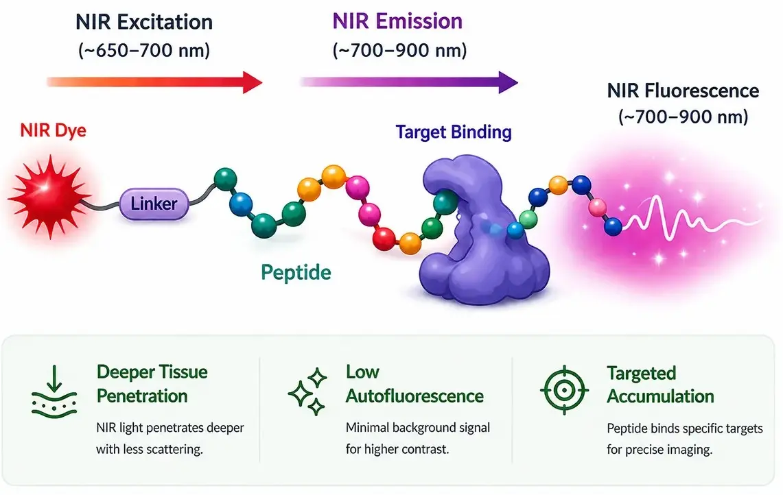
Core architecture: NIR Dye — Linker — Peptide. The dye provides fluorescence; the linker manages spacing, solubility, and steric accessibility; the peptide directs the conjugate toward a biological target.

Key Features of NIR Peptide Conjugates

Targeted Signal

Peptide ligands can direct fluorescence toward receptors, enzymes, tumor markers, or tissue-specific targets.

Low Autofluorescence

NIR emission reduces background from biological samples compared with many visible-range fluorophores.

Deeper Tissue Imaging

Longer wavelengths can improve tissue penetration and reduce scattering in optical imaging workflows.

Flexible Conjugation

Dyes can be attached through terminal, side-chain, click, maleimide, NHS ester, or project-specific chemistries.

How NIR Peptide Conjugates Work

NIR peptide conjugates enable targeted imaging through near-infrared dye excitation, peptide-mediated binding, and fluorescence signal detection within biological systems.

How NIR peptide conjugates work showing excitation, emission, targeting, and fluorescence detection
NIR peptide conjugate workflow: excitation, emission, targeting, and signal detection.

Key Steps

  • Excitation: NIR light (~650–700 nm) activates the dye
  • Emission: Dye emits fluorescence (~700–900 nm)
  • Targeting: Peptide binds to specific cells or biomolecules
  • Detection: Signal is detected within tissues

Typical Optical Window

Excitation range ~650–700 nm
Emission range ~700–900 nm
Detection Fluorescence imaging systems
Targeting unit Peptide ligand or binding motif
Format Dye – Linker – Peptide

NIR Dye Options & Spectral Reference

Common near-infrared dyes used in peptide conjugates with their typical excitation and emission ranges, along with representative applications.

Dye class Excitation (nm) Emission (nm) Typical applications
Cy5 ~640–650 ~660–680 Far-red imaging, microscopy, receptor binding assays
Cy5.5 ~670–690 ~690–710 Tissue imaging, optical biodistribution studies, red-shifted fluorescence workflows
Alexa Fluor 680 / 700 ~680–700 ~700–720 Scanner-based detection, microscopy, fluorescence assays
Cy7 / Alexa Fluor 750 ~730–760 ~760–790 In vivo optical imaging, deeper tissue visualization, low-background studies
IRDye 800-class ~770–790 ~790–820 Multiplex imaging, biodistribution analysis, high-sensitivity detection
Custom NIR dyes Variable Variable Project-specific conjugates, advanced platforms, and instrument-matched dye selection
Selection tip: Choose dyes based on instrument excitation source, emission filters, detector sensitivity, sample background, dye hydrophobicity, and multiplex compatibility.

Peptide Labeling Sites and Conjugation Handles

NIR dye placement can affect target binding, solubility, stability, and fluorescence performance. The best site is usually away from the peptide’s binding motif or active pharmacophore.

Labeling position Description Typical use patterns
N-terminal labeling NIR dye attached at the N-terminus through an amine-reactive or pre-installed linker strategy. Common when the N-terminus is not required for target binding.
C-terminal labeling Dye installed at or near the C-terminus through a terminal handle or linker. Useful when the C-terminal region is accessible and does not disrupt activity.
Lysine side-chain labeling Dye attached to an epsilon-amine on a selected lysine residue. Good for internal or side-chain labeling when position-specific placement is planned.
Cysteine / maleimide labeling Dye attached to a free thiol or protected cysteine handle. Useful for site-specific labeling, especially when only one reactive cysteine is available.
Click chemistry handles Azide, alkyne, DBCO, or related orthogonal groups used for selective dye conjugation. Useful for complex peptides or late-stage conjugation strategies.
Spacer-assisted labeling PEG, Ahx, beta-alanine, or other spacers separate the dye from the peptide. Helps reduce steric interference, aggregation, or dye-driven loss of binding.

Design Considerations for NIR Peptide Conjugates

Preserve peptide activity

Place the dye away from residues required for binding, receptor recognition, enzyme processing, or cell penetration.

  • Identify binding-critical residues
  • Avoid blocking active motifs
  • Use spacers if steric interference is likely

Control hydrophobicity

NIR dyes are often hydrophobic and can reduce solubility or increase aggregation unless linker and formulation are considered.

  • Consider PEG or hydrophilic spacers
  • Review sequence hydrophobicity
  • Plan storage and handling conditions

Match the instrument

Dye choice should match the actual excitation and emission settings of the microscope, scanner, or in vivo imager.

  • Confirm laser/LED excitation
  • Confirm emission filter set
  • Plan multiplex channel separation
Fastest design review: Send the peptide sequence, intended target, residues that must remain unmodified, preferred dye or wavelength, labeling site, linker preference, quantity, purity target, and intended imaging platform.

Applications of NIR Peptide Conjugates

Tumor Imaging

  • Targeted visualization of tumor-associated receptors
  • Preclinical imaging and localization studies
  • Fluorescence-guided research workflows

Receptor Imaging

  • Ligand-receptor binding studies
  • Target expression and distribution analysis
  • Competitive binding and uptake assays

Biodistribution Studies

  • Track peptide localization over time
  • Compare linker or dye variants
  • Evaluate tissue retention and clearance

Cell-Penetrating Peptides

  • Monitor cellular uptake
  • Optimize peptide delivery systems
  • Study intracellular localization patterns

Enzyme Activity Imaging

  • Fluorescent peptide substrates
  • Protease-sensitive linker concepts
  • Activity-linked signal readouts

Drug Delivery Research

  • Peptide-drug conjugate tracking
  • Payload delivery route evaluation
  • Target engagement and accumulation studies

Product Specification Guide

Parameter Typical options Notes
Conjugate format NIR Dye — Linker — Peptide Architecture can be customized around the binding motif and detection goal.
Peptide length Short motifs to longer custom peptides Feasibility depends on sequence, hydrophobicity, modifications, and folding requirements.
Labeling site N-terminus, C-terminus, lysine, cysteine, click handle, or custom handle Site-specific design is recommended for active peptides.
Linker options PEG, Ahx, beta-alanine, flexible alkyl spacers, cleavable linkers, or custom spacers Linkers help tune spacing, solubility, and target accessibility.
Dye options Cy5, Cy5.5, Cy7, Alexa Fluor-class dyes, IRDye-class dyes, or project-specific NIR dyes Availability and suitability are project-dependent.
Purification HPLC purification; purity target matched to application Higher purity may be preferred for sensitive imaging workflows.
QC Analytical HPLC, LC-MS or MS, optional UV-Vis/fluorescence confirmation Final QC package depends on conjugate chemistry and application requirements.
Storage Protect from light; usually frozen or lyophilized storage when appropriate Handling recommendations depend on dye and peptide chemistry.

Workflow: From Design to Delivery

1

Define the construct

Confirm peptide sequence, target, dye region, labeling site, linker, and quantity.

2

Synthesize peptide

Build the peptide with required handles, protecting groups, or orthogonal chemistry.

3

Conjugate dye

Attach the NIR fluorophore using the selected terminal, side-chain, or click chemistry route.

4

Purify and release

Purify the final conjugate and provide analytical documentation for project use.

Quote checklist: sequence, dye or wavelength range, labeling site, linker preference, quantity, purity target, QC needs, and intended use in cells, tissue, animal imaging, receptor binding, or analytical detection.

QC & Deliverables

Analytical Confirmation

  • Analytical HPLC or UPLC profile
  • Mass confirmation when applicable
  • Conjugate identity and modification summary

Fluorescence-Ready Handling

  • Light-protected handling recommendations
  • Lyophilized or solution format when appropriate
  • Storage guidance based on dye and peptide chemistry

Documentation

  • Certificate of Analysis
  • Purity and identity data
  • Optional UV-Vis or fluorescence documentation

FAQ

What are near-infrared peptide conjugates?

They are peptides covalently linked to NIR fluorescent dyes for targeted fluorescence imaging, receptor binding studies, biodistribution, and optical detection workflows.

Why are NIR dyes useful for peptide imaging?

NIR dyes can reduce biological autofluorescence and improve tissue penetration compared with many visible dyes, which helps increase signal-to-background contrast.

Where should the dye be attached?

The dye should usually be placed away from the active binding motif. Common positions include the N-terminus, C-terminus, lysine side chain, cysteine side chain, or a click handle.

Can you add a linker between the dye and peptide?

Yes. PEG, Ahx, beta-alanine, flexible spacers, or cleavable linkers can be used to improve spacing, reduce steric interference, or tune solubility.

Which dyes are commonly used?

Common options include Cy5, Cy5.5, Cy7, Alexa Fluor 680/750, IRDye 680/800, and other project-specific NIR fluorophores.

What information is needed for a quote?

Send the peptide sequence, preferred dye or wavelength, labeling site, linker preference, quantity, purity target, QC requirements, and intended imaging or binding application.

Contact & Quote Request

For the fastest quote on custom NIR peptide conjugates, share the peptide sequence, desired dye or wavelength range, labeling position, linker preference, quantity, purification target, analytical requirements, and intended imaging or detection workflow.

Fast quote checklist

  • Peptide sequence and target/application
  • Preferred NIR dye or emission range
  • N-terminal, C-terminal, lysine, cysteine, or click-handle labeling
  • Quantity, purity target, and QC/documentation needs

Fastest path

Why Choose Bio-Synthesis

Trusted by biotech leaders worldwide for over 45+ years of delivering high quality, fast and scalable synthetic biology solutions.